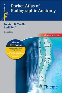
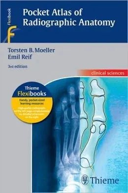
Build confidence interpreting conventional diagnostic radiographs with this pocket atlas designed for fast, practical reference. Two expert radiologists present normal radiographic anatomy in a consistent two-page format, pairing each standard image with a clearly labeled schematic for quick review.
Key benefits
- 177 radiographic studies with multiple views to support interpretation of every basic anatomic structure
- High-resolution radiographs presented beside explanatory drawings to strengthen comprehension
- Seven examinations new to this edition, including a trans-scapular Y view of the shoulder and 45 external and internal rotation views of the knee
- Easy-to-use, consistent layout that helps you identify and rapidly revisit key anatomic information
- Includes standard radiographs for 170 radiographic examinations and their different projections
Who this is for
- Medical students who need a dependable, portable anatomy reference for radiographic interpretation
- Radiology residents looking for quick normal anatomy review during training
- Radiological technologists who benefit from a clear, projection-based approach to commonly performed studies
Details
- Title: Pocket Atlas of Radiographic Anatomy, 3rd Edition (ORIGINAL PDF from Publisher)
- Edition: 3
- Authors: Torsten B. Möller, Emil Reif
- Publisher: Thieme
- Published Date: 2000
- Pages: 374
- ISBN: 9781604061307, 1604061308
- Brand / Publisher: Orthopedics Library
Get the pocket atlas you can rely on for rapid, normal radiographic anatomy review. Add this 3rd edition from Orthopedics Library to your collection today.